Are Chromosomes Visible During Mitosis? Explained & Answered
Are chromosomes visible during mitosis?
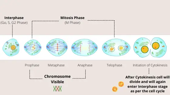
Yes, chromosomes are visible during the mitosis phase (M phase) of the cell cycle and cell division. We can see chromosomes during the mitosis phase because as the mitosis begins the initiation of condensation of chromosomal materials occurs and this causes the chromosomes to get untangled and thickened and thus become visible under a microscope.
DNA replication occurs during the S phase (Synthesis phase) of the interphase stage which can take anywhere between 5 to 6 hours to complete. During this phase, the chromosomes remain as semi-condensed chromatin fibres.
And, as the S phase gets over and then following the G2 phase, the process of mitosis (M phase) starts which lasts for about 1 hour approximately.
Prophase, Metaphase, Anaphase, and Telophase are the 4 serial stages of the cell cycle that occur one after another during this mitosis phase.
And starting from the prophase stage to the anaphase stage of mitosis, the chromosomes become visible under a microscope which is focused with a 40x objective lens.
There are two important SMC (Structural Maintenance of Chromosomes) proteins that play a key role in chromosome condensation, organization, and dynamics during the cell cycle.
These two prominent proteins are called Condensin and Cohesin. And that, these proteins are synthesized during the S phase of Interphase stage in the cell cycle.
Condensin and Cohensin play a primary role in chromosome assembly and segregation from the chromosomal materials in eukaryotes by causing the condensation of chromosomal materials during the mitosis phase.
Condensin does so by promoting the condensation of chromosomal materials by linking two distant segments of single chromatids.
On the other hand, Cohesin promotes the holding of the two sister chromatids that were linked by condensin in order to keep the sister chromatids connected with each other to form the chromosome.
So, thus it can be also stated that due to the presence and functions of these two proteins the chromosome assembly and segregation occurs from the loosely packed chromatin fibers leading to the visibility of chromosomes during the mitosis phase (M phase) of the cell cycle.

Why are chromosomes only visible during mitosis and not during interphase?
During the interphase stage of cell division, the chromosomal materials don’t exist as chromosomes. In fact, they exist as thin, lengthy, and loosely packed chromatin fibers which are simply a complex of DNA, proteins, and RNA found in eukaryotic cells.
Chromosomes are visible during mitosis because those thin, lengthy, and loosely packed chromatin fibers now start to get condensed and compactly arranged to form two chromatids being attached to one another as a single chromosome.
The role of the two multi-dimensional proteins named Cohesin and Condensin plays a key role in causing the chromatin fibres (also called chromosomal materials) to take their compact shape and become visible under the microscope.
The chromatin fibres look like a “beads on string” structure. It’s due to the presence of nucleosomes that make such a structure to the chromosomes.
The primary function of chromatin fibres is to efficiently package DNA and proteins into a small volume to fit that DNA and proteins into the nucleus of a cell and protect the DNA structure and sequence.
And so yes, a chromosome is made up of chromatin fibre. It’s actually the chromosomes that are finely packaged and compressed structure of chromatin fibres.
These chromatin fibres remain inside the nucleus when it is not the mitosis phase of cell cycle.
And so, when inside the nucleus they don’t have easily identified features when viewed under a light microscope due to its very loose packaging, thus showing no visibility at all.
So, When are chromosomes visible in mitosis?
It is strictly during the Prophase, Metaphase, and Anaphase stage of mitosis when the chromosomes are perfectly visible under a microscope with higher magnification of 40x objective.
Mitosis includes both karyokinesis and cytokinesis. During karyokinesis, the nuclear division takes place, whereas during cytokinesis cytoplasmic division leading to the division of a single whole cell into two different cells takes place.
Karyokinesis can be seen during all of the four stages of mitosis i.e. during Prophase, Metaphase, Anaphase, and Telophase.
So, it can be also stated that not in all phases of karyokines chromosomes can be seen, as chromosomes are not visible during the Telophase stage. And also that, during cytokinesis no chromosomes are visible at all.
During telophase stage the cytoplasm division leading to cell division has not occurred but the nucleus and the nuclear membrane has formed with the loosely packed chromatin fibers inside. So, chrosomes are not visible during telophase.
Also during Cytokinesis that falls just after Telophase of mitosis in which the nucleus’s genetic material remains unchanged and remains as it was during telophase with the loosely packed chromatin fibers inside. So, chromosomes are also not visible during cytokinesis.
Interphase is the stage between two successive mitosis and it is the time when the cells contain a nucleus with a distinctive nuclear membrane. During this phase the nucleus contains semi-condensed chromatin fibres which aren’t visible at all under a microscope as clear-cut chromosomes.
The difference between semi-condensed chromatin fibres, full condensed chromatin fibres, and loosely packed chromatin fibers is that the semi-condensed chromatin fibres don’t take the structure of chromosomes and so they look like a thick patch of concentrated area in the nucleus of the cell called the nucleolus.
While, full condensed chromatin fibres take the fine shape of chromosomes with having two distinct chromatids in each of the chromosome.
On the other case, loosely packed chromatin fibers are not visible as chromosomes at all because they are so small and at the simplest level they are just double-stranded helical structures of DNA along with histone proteins.
And so, by understanding this difference we can also state that semi-condensed chromatin fibres are not visible as chromosomes which is during the Interphase stage.
And also that, full condensed chromatin fibres are visible as chromosomes which is during the Prophase, Metaphase, and Anaphase stages of mitosis.
To make it easy to understand just go through the below points
- The cell during the Interphase stage will have semi-condensed chromatin fibers. So, not visible as true chromosomes, but visible as a thick dark patch inside the nucleus.
- The cell during the Prohase, Metaphase, and Anaphase stages of Mitosis will have fully condensed chromatin fibres. So, visible as true chromosomes having chomatids.
- The cell during the Telophase and Cytokinesis stage will have loosely packed chromatin fibres. So, chromosomes are not visible at all.
Are chromosomes only visible during cell division?
Yes, chromosomes are only visible during cell division and not at all times. The visibility of chromosomes lies in the phenomenon of DNA packaging which states that the chromosomes are actually formed due to the coiling and supercoiling of chromatin fibers in certain stages of the cell cycle.
In other times i.e when the cell is not dividing at all, the DNA will simply lie in the nucleus as loosely packed chromatin fibres, which actually are now uncoiled DNA.
But, during certain stages of the cell cycle i.e. during prophase, metaphase, and anaphase time these chromatin fibres will coil and supercoil themselves to take the shape of fully condensed chromatin fibres known as chromosomes. This will now be supercoiled DNA.
Each Chromosome is up made of:
- A single DNA molecule: A DNA molecule is referred to the whole long DNA strand that coils and supercoils itself to take the shape of a chromosome during cell division.
- Histone Proteins: Generally, 8 Histone proteins (also called a histone octamer) form a bead-like structure around which the DNA takes 1.7 turns to form a nucleosome.
So, it is that’s why said that during the cell division, chromosomes are highly condensed with the DNA being supercoiled, and so become visible under a light microscope as clear-cut chromosomes with their respective chromatids.
