Gram-Positive and Gram-Negative Bacteria: Differences in Structure, Function, and Antibiotic Resistance
Table of Contents
I. Introduction to Bacterial Classification
Bacterial classification is vital in microbiology as it informs our understanding of their diverse structures, functions, and responses to environmental challenges, particularly antibiotics. This process of classification goes beyond simply grouping bacteria; it also provides critical insight into their ecological roles and interactions with other organisms. At the core of this classification are the distinctions between Gram-positive and Gram-negative bacteria, which are primarily based on their unique cell wall compositions. Gram-positive bacteria possess a thick peptidoglycan layer that allows them to retain crystal violet dye during the Gram staining process, resulting in a characteristic blue or purple appearance under the microscope. In contrast, Gram-negative bacteria are defined by their thinner peptidoglycan layer, which is sandwiched between an outer membrane and the periplasmic space, leading to a pink coloration upon staining. This structural difference is not merely aesthetic; it significantly impacts the bacteria’s susceptibility to antibiotics. For instance, the outer membrane found in Gram-negative bacteria acts as a barrier to many types of antibiotics, making them inherently more resistant to these treatments compared to their Gram-positive counterparts. Additionally, Gram-negative bacteria often exhibit specific resistance mechanisms, such as efflux pumps, which actively expel harmful substances from their cells, further complicating treatment strategies, as illustrated in [citeX]. Thus, understanding these distinctions is imperative for developing effective antimicrobial strategies and addressing the challenges posed by antibiotic resistance. This classification framework not only aids in clinical diagnosis and treatment but also helps researchers devise new methods to combat bacterial infections and improve public health outcomes.
| Classification | Cell Wall Structure | Examples | Antibiotic Resistance | Typical Diseases |
| Gram-Positive Bacteria | Thick peptidoglycan layer | Staphylococcus aureus, Streptococcus pneumoniae | Generally sensitive to penicillin, but resistance is increasing. | Skin infections, pneumonia, endocarditis |
| Gram-Negative Bacteria | Thin peptidoglycan layer plus outer membrane | Escherichia coli, Salmonella enterica | Often resistant to penicillin; more likely to have multi-drug resistance. | Urinary tract infections, gastrointestinal infections |
| Bacterial Classification Summary | Varies | Includes both Gram-positive and Gram-negative bacteria | Resistance patterns differ significantly between groups. | Wide range of infections caused by different species |
Bacterial Classification Overview
Here is a detailed comparison of Gram-positive and Gram-negative bacteria, highlighting 30 key differences. This table summarizes the fundamental structural, chemical, and physiological differences between Gram-positive and Gram-negative bacteria, which influence their function, antibiotic resistance, and ecological roles.
| Feature | Gram-Positive Bacteria | Gram-Negative Bacteria |
|---|---|---|
| 1. Cell Wall Composition | Thick peptidoglycan layer (20–80 nm) | Thin peptidoglycan layer (2–7 nm) |
| 2. Outer Membrane | Absent | Present |
| 3. Peptidoglycan Percentage | 60–90% of the cell wall | 10–20% of the cell wall |
| 4. Lipopolysaccharides (LPS) | Absent | Present in the outer membrane |
| 5. Teichoic Acids | Present (provides rigidity and antigenic properties) | Absent |
| 6. Periplasmic Space | Small or absent | Large and well-defined |
| 7. Gram Staining | Retains crystal violet stain and appears purple | Does not retain crystal violet, takes safranin stain, appearing pink/red |
| 8. Cell Wall Rigidity | More rigid due to thick peptidoglycan | Less rigid due to thin peptidoglycan |
| 9. Resistance to Physical Disruption | More resistant due to thick wall | Less resistant due to thinner wall |
| 10. Susceptibility to Lysozyme | More susceptible (lysozyme digests peptidoglycan) | Less susceptible due to outer membrane protection |
| 11. Sensitivity to Penicillin | More sensitive (Penicillin inhibits peptidoglycan synthesis) | Less sensitive (Outer membrane blocks many antibiotics) |
| 12. Lipid Content in Cell Wall | Low lipid content | High lipid content due to outer membrane |
| 13. Endotoxins | Absent | Present (LPS contains endotoxin, mainly Lipid A) |
| 14. Exotoxin Production | Mainly exotoxin producers | Some produce exotoxins, but mainly endotoxins |
| 15. Flagella Structure | 2 basal body rings in flagella | 4 basal body rings in flagella |
| 16. Pili/Fimbriae | Less common | More common |
| 17. Spore Formation | Some species form endospores (e.g., Bacillus, Clostridium) | Rarely form endospores |
| 18. Resistance to Drying | More resistant due to thick peptidoglycan | Less resistant due to thin wall and high lipid content |
| 19. Susceptibility to Complement System | Less susceptible | More susceptible due to outer membrane permeability |
| 20. Ribosomes (70S) | 70S ribosomes (common to all bacteria) | 70S ribosomes (common to all bacteria) |
| 21. Plasmids | Present in many species | Present in many species |
| 22. Pathogenicity | Many species are pathogenic (e.g., Streptococcus, Staphylococcus) | Many species are pathogenic (e.g., Escherichia coli, Salmonella) |
| 23. Nutrient Transport Mechanism | Simple due to permeability of thick peptidoglycan | Complex due to selective outer membrane |
| 24. Lipoproteins | Absent or rare | Present, anchoring the outer membrane to peptidoglycan |
| 25. Sensitivity to Alcohol and Detergents | More resistant due to thick peptidoglycan | More sensitive due to outer membrane disruption |
| 26. Oxidation Stress Tolerance | More resistant to oxidative damage | Less resistant to oxidative stress |
| 27. Genetic Variability | Generally lower horizontal gene transfer rates | Higher horizontal gene transfer due to conjugation mechanisms |
| 28. Representative Genera | Bacillus, Clostridium, Staphylococcus, Streptococcus | Escherichia, Salmonella, Pseudomonas, Neisseria |
| 29. Growth on Selective Media | Easier to grow on simple media | Often requires enriched/selective media |
| 30. Role in Biotechnology | Widely used in industrial fermentation (antibiotics, enzymes) | Important in genetic engineering and antibiotic resistance studies |
A. Why Gram Staining is Important in Microbiology
Gram staining is a fundamental technique in microbiology that offers critical insights into the structural and functional differences between Gram-positive and Gram-negative bacteria, which significantly influences their susceptibility to antibiotics. By utilizing this staining method, microbiologists can differentiate bacteria based on the characteristics of their cell walls, which are categorized by the presence of thick peptidoglycan layers in Gram-positive organisms and the complex outer membrane structures found in their Gram-negative counterparts. This crucial distinction aids in the identification of various bacterial species, thereby guiding clinicians in making informed decisions about appropriate antibiotic selections and treatment strategies tailored to combat infections effectively. For instance, the enhanced permeability of Gram-negative bacteria, as illustrated in [citeX], portrays how their unique outer membrane can hinder the efficacy of certain antibiotics, rendering them less susceptible to standard treatments. Furthermore, understanding these differences in cell structure is essential for research focused on combating antibiotic resistance, as highlighted by the resistance mechanisms depicted in [extractedKnowledgeX]. The implications of this knowledge extend beyond mere classification; they also inform the development of new therapeutic approaches. Through Gram staining, microbiologists can not only classify bacteria but also devise targeted strategies to manage infections effectively, addressing the ever-evolving challenges posed by resistant strains. This technique proves vital in clinical and research settings alike, as it allows for the identification of pathogenic organisms in a timely manner, ultimately improving patient outcomes and guiding public health interventions. Therefore, Gram staining remains a cornerstone of microbiological diagnostics and treatment planning, continuing to significantly contribute to advancements in medical microbiology.

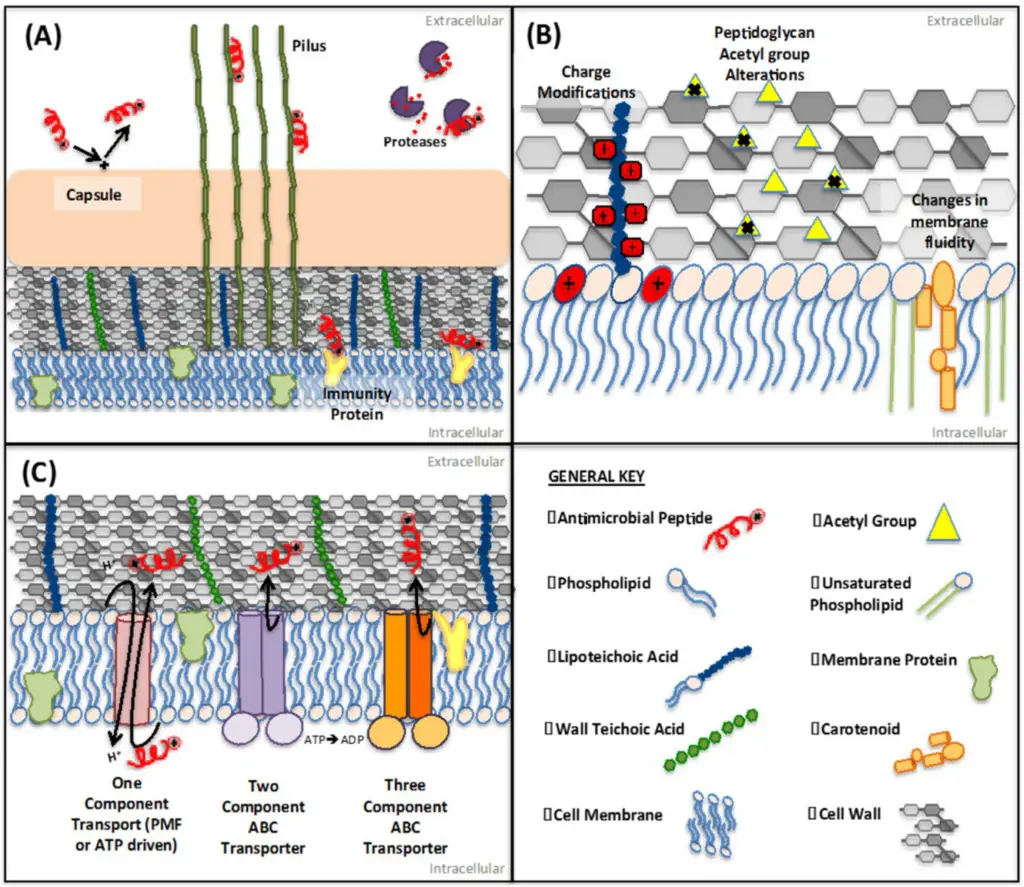
IMAGE – Schematic representation of bacterial membrane structures and transport mechanisms. (The image provides a detailed schematic representation of bacterial membrane structures and interactions. Panel (A) illustrates the components of the bacterial envelope, including the capsule, pili, proteases, and immunity proteins, emphasizing their roles in interaction with external factors. Panel (B) focuses on peptidoglycan alterations, charge modifications, and changes in membrane fluidity, which are significant for understanding bacterial resistance mechanisms. Panel (C) depicts various transport mechanisms across the bacterial membrane, including one-component transport, two-component ABC transporters, and three-component ABC transporters. Accompanying this, a general key defines the various components represented in the diagrams, such as antimicrobial peptides, phospholipids, and membrane proteins, which are crucial for comprehending bacterial physiology and pathogenicity.)
| Aspect | Description |
| Identifying Bacteria | Gram staining helps differentiate between Gram-positive and Gram-negative bacteria, allowing for accurate identification. |
| Guiding Treatment | Determining the Gram status of bacteria influences antibiotic selection and treatment strategies. |
| Understanding Pathogenic Mechanisms | Gram staining aids in studying the structural differences in bacteria, which can inform understanding of their pathogenicity. |
| Research Applications | It is a fundamental technique used in research to classify bacteria quickly and efficiently. |
| Clinical Diagnostics | Gram staining is routinely used in clinical laboratories to provide rapid results for infection diagnosis. |
Gram Staining Importance in Microbiology
B. How the Cell Wall Defines Bacterial Properties
The composition and structure of bacterial cell walls are fundamental in defining their physical properties, functionality, and responses to environmental stresses, including antibiotic treatment. Gram-positive bacteria are characterized by a thick peptidoglycan layer that not only provides structural integrity but also plays a crucial role in retaining crystal violet dye during Gram staining. This characteristic feature significantly influences their susceptibility to certain antibiotics, such as penicillin, which specifically target peptidoglycan synthesis. In contrast, Gram-negative bacteria possess a thinner peptidoglycan layer sandwiched between two membranes, which complicates the landscape of antibiotic treatment due to the presence of an outer membrane that acts as a formidable barrier. This distinction underpins their differential antibiotic resistance mechanisms, including the presence of efflux pumps that actively expel harmful substances, and porins that selectively regulate the passage of essential molecules and ions into the cell. These unique features of the bacterial cell wall not only determine the effectiveness of antibiotic therapy but also impact other aspects of bacterial physiology, such as cell shape, rigidity, and the ability to withstand osmotic pressure. Understanding these structural variances emphasizes the importance of the cell wall in determining the bacterium’s survival strategies and interactions with therapeutic agents, as illustrated in [citeX]. The interplay between cell wall composition and environmental factors reveals a complex relationship that is critical for devising effective treatment strategies against bacterial infections, demonstrating how the cell wall is integral not only to structural integrity but also to overall bacterial adaptability and resilience.
| Type | Cell Wall Composition | Outer Membrane | Teichoic Acids | Example Antibiotics Effective | Antibiotic Resistance Rate (%) |
| Gram-Positive | Thick peptidoglycan layer | Absent | Present | Penicillin, Vancomycin | 10 |
| Gram-Negative | Thin peptidoglycan layer | Present | Absent | Ciprofloxacin, Gentamicin | 30 |
| Gram-Positive (Staphylococcus aureus) | Thick peptidoglycan layer | Absent | Present | Methicillin | 40 |
| Gram-Negative (Escherichia coli) | Thin peptidoglycan layer | Present | Absent | Ampicillin | 25 |
Bacterial Cell Wall Characteristics and Antibiotic Resistance
II. Characteristics of Gram-Positive Bacteria
The characteristics of Gram-positive bacteria are defined primarily by their thick peptidoglycan layer, which constitutes a significant portion of their cell wall structure. This robust layer not only provides structural integrity but also plays a crucial role in the retention of the crystal violet stain used in Gram staining, a fundamental technique for bacterial classification that helps differentiate between various types of bacteria. Unique to Gram-positive bacteria are teichoic acids, which are embedded within the peptidoglycan and contribute to the overall rigidity and strength of the cell wall while potentially serving as receptors for bacteriophages, thus influencing their interactions with viruses. Unlike their Gram-negative counterparts, Gram-positive bacteria lack an outer membrane, a distinctive feature that significantly influences their susceptibility to certain antibiotics and detergents. This absence of an outer membrane means that Gram-positive bacteria are often more exposed to environmental threats, making them more vulnerable to antimicrobial agents; therefore, their simplistic structure allows for the direct interaction of these agents with the cell wall, enhancing their vulnerability to compounds like penicillin and other beta-lactam antibiotics. The spatial arrangement of these various components is exemplified in numerous studies, which succinctly illustrate the defining features of Gram-positive bacterial architecture and underlines their functional implications in microbiological research. Understanding these characteristics is vital not only for classification purposes but also for developing effective treatments against bacterial infections caused by these organisms, which are of significant clinical concern in both human and veterinary medicine.
| Characteristic | Description | Presence of Teichoic Acids | Gram Staining Result | Examples |
| Cell Wall Structure | Thick peptidoglycan layer. | Present | Purple | Staphylococcus, Streptococcus |
| Cell Membrane Structure | Single phospholipid bilayer beneath the peptidoglycan layer. | Absent | Purple | Bacillus, Clostridium |
| Antibiotic Sensitivity | Generally more susceptible to antibiotics targeting peptidoglycan synthesis. | Some may develop resistance through mutations or enzymatic degradation. | Purple | Enterococcus, Listeria |
| Endospore Formation | Some genera, like Bacillus and Clostridium, can form highly resistant endospores under stress. | Variable | Purple | Bacillus anthracis, Clostridium difficile |
Characteristics of Gram-Positive Bacteria
A. Structure of Gram-Positive Cell Walls
The structure of Gram-positive cell walls is a defining characteristic that distinguishes them from Gram-negative bacteria and contributes significantly to their biological functions and interactions with antibiotics. Composed primarily of a thick layer of peptidoglycan, which can be up to 90% of the cell wall, Gram-positive bacteria exhibit a robust architecture that not only provides structural integrity but also offers protection against various environmental stressors such as physical forces and osmotic pressure. This thick peptidoglycan layer is crucial for maintaining the shape of the bacteria and preventing lysis in hypotonic conditions. In addition to peptidoglycan, the presence of teichoic and lipoteichoic acids plays a crucial role in maintaining wall rigidity, serving as scaffolding, and contributing to cell signaling processes that facilitate interactions with their surroundings. These acids can also mediate adhesion to surfaces and evasion of immune responses. Unlike their Gram-negative counterparts, which possess an outer membrane, these layers of peptidoglycan and acids directly interface with the external environment, influencing susceptibility to various antibiotics and the ability of host immune responses to target the bacteria effectively. As depicted in [extractedKnowledgeX], the unique composition and arrangement of Gram-positive cell walls are critical for understanding their functionality, especially in the context of antibiotic resistance. This highlights important avenues for developing targeted antimicrobial strategies against these resilient pathogens, ensuring that researchers and clinicians can formulate effective treatments while addressing public health challenges associated with resistant strains.
| Bacteria Type | Peptidoglycan Layer Thickness (nm) | Teichoic Acids Present | Outer Membrane | Common Examples |
| Gram-Positive | 20 | Yes | No | Staphylococcus aureus, Streptococcus pneumoniae |
| Gram-Negative | 5 | No | Yes | Escherichia coli, Salmonella enterica |
Comparison of Cell Wall Structure in Gram-Positive and Gram-Negative Bacteria
B. Common Gram-Positive Bacteria (Staphylococcus, Streptococcus)
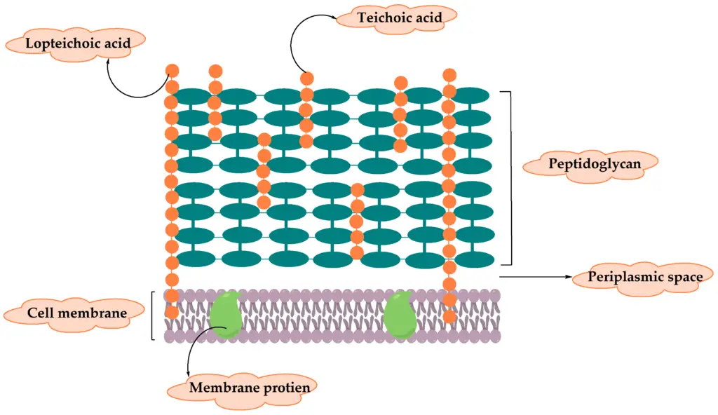
Among the well-known Gram-positive bacteria, Staphylococcus and Streptococcus play significant roles in human health and disease, influencing both the field of microbiology and clinical practice extensively. Staphylococcus, characterized by its clustered spherical shape, is frequently found residing on the skin and mucous membranes of humans, making it a common inhabitant of the body. However, it can transition into a pathogenic state under certain conditions, leading to various types of infections, including skin infections, pneumonia, and sepsis, which can pose serious health risks, especially in immunocompromised individuals. Streptococcus, in stark contrast, exists in chains and is particularly notorious for its involvement in pharyngitis, commonly known as strep throat, and pneumonia, as well as in more severe infections such as rheumatic fever, which can have long-lasting effects on heart health. Both genera exhibit notable discrepancies in their structural components, particularly in their cell wall composition and associated properties, which contribute to their differing mechanisms of antibiotic resistance. Understanding these distinctions is imperative for developing targeted therapeutic strategies that can effectively combat infections caused by these bacteria. For instance, the structural details of bacterial cell walls illustrated in [citeX] can enhance the comprehension of how these bacteria retain their integrity and function, directly informing the approach to antibiotic treatment in clinical settings while also guiding the creation of new antimicrobial agents. Such knowledge is crucial not only for effective treatment but also for preventing the spread of antibiotic-resistant strains that pose increasing challenges to public health systems worldwide.

Image : Structural Components of Bacterial Cell Wall (The diagram illustrates the structural components of a bacterial cell wall, emphasizing elements such as lipoteichoic acid, teichoic acid, peptidoglycan, periplasmic space, cell membrane, and membrane proteins. It visually represents the hierarchical organization of these components, with annotations indicating their specific roles and relationships within the structure. This representation can facilitate understanding of bacterial cell wall composition and function in microbiology.)
| Bacteria | Characteristics | Common Infections | Antibiotic Resistance |
| Staphylococcus aureus | Cocci shape, clusters; produces toxins and enzymes. | Skin infections, pneumonia, food poisoning. | MRSA is a variant resistant to methicillin. |
| Streptococcus pneumoniae | Cocci shape, chains; encapsulated. | Pneumonia, meningitis, sinusitis. | Some strains show penicillin resistance. |
| Streptococcus pyogenes | Cocci shape, chains; beta-hemolytic. | Strep throat, skin infections, rheumatic fever. | Generally susceptible, but rising resistance reported. |
Common Gram-Positive Bacteria
C. Antibiotic Sensitivity and Resistance
The phenomenon of antibiotic sensitivity and resistance is critically influenced by the structural differences between Gram-positive and Gram-negative bacteria. Gram-positive bacteria, characterized by a thick peptidoglycan layer, often exhibit inherently high sensitivity to antibiotics such as penicillin, which specifically target the critical process of cell wall synthesis. This characteristic makes them more vulnerable to certain antibiotic classes, allowing for successful treatment options in many infections caused by these organisms. In contrast, Gram-negative bacteria possess an outer membrane that acts as an additional barrier, complicating the penetration of antimicrobial agents and often rendering them resistant to many conventional antibiotics, including penicillins and cephalosporins. This outer membrane not only hinders drug entry but also contains porins that selectively allow the passage of certain molecules while obstructing others. Mechanisms such as the production of beta-lactamases, which efficiently degrade beta-lactam antibiotics, are a prominent means of resistance observed in both bacterial groups; however, their prevalence and effectiveness can vary significantly between species. Furthermore, the presence of efflux pumps in Gram-negative bacteria significantly enhances their ability to expel various antibiotics before they can exert their intended effects on the bacterial cell. These efflux pumps contribute to multi-drug resistance, allowing for survival in the presence of multiple antibiotics. Understanding these intricate relationships and mechanisms of resistance is essential for developing targeted therapeutic strategies against resistant bacterial strains, as illustrated in [citeX], which effectively underscores the complexity of bacterial membrane interactions and resistance mechanisms. Working towards solutions that address these challenges remains a high priority in the field of infectious diseases.
| Bacteria Type | Common Antibiotics Used | Resistance Rate | Notes |
| Gram-Positive | Penicillin, Vancomycin, Daptomycin | 5-15% | Generally more susceptible to antibiotics but emergence of resistant strains observed. |
| Gram-Negative | Ciprofloxacin, Meropenem, Gentamicin | 30-60% | Higher resistance rates due to efflux pumps and outer membrane barrier. |
| Enterobacteriaceae | Ceftriaxone, Amikacin, Imipenem | 50-70% | Significant increases in resistance due to extended-spectrum beta-lactamases (ESBLs). |
| Staphylococcus aureus | Methicillin, Clindamycin, Linezolid | 20-50% | Methicillin-resistant Staphylococcus aureus (MRSA) is a major concern. |
| Pseudomonas aeruginosa | Piperacillin-tazobactam, Ceftazidime, Polymyxins | 30-40% | Known for its multidrug resistance patterns. |
Antibiotic Sensitivity and Resistance in Gram-Positive and Gram-Negative Bacteria
III. Characteristics of Gram-Negative Bacteria
Gram-negative bacteria are distinguished by their complex cell structure, which prominently features an outer membrane composed of lipopolysaccharides (LPS) and phospholipids. This outer membrane serves as a formidable barrier against environmental threats, including antibiotics, while simultaneously protecting the bacteria from harmful substances such as detergents, dyes, and bile salts, thereby enhancing their survival in diverse environments. The periplasmic space, located between the outer membrane and the inner cytoplasmic membrane, houses a variety of enzymes and proteins that further facilitate nutrient transport and defend against oxidative stress as well as enzymatic degradation. Notably, the presence of porins in the outer membrane permits the selective passage of small molecules such as nutrients and waste products; however, this selective permeability also contributes to antibiotic resistance by limiting the entry of various antimicrobial agents that might otherwise disrupt bacterial function. The structural integrity provided by the peptidoglycan layer, although thinner than that of Gram-positive bacteria, plays a critical role in maintaining cell shape and rigidity while also offering some measure of protection against external stresses. These characteristics underscore the remarkable adaptability of Gram-negative bacteria, particularly in their ability to acquire and develop resistance mechanisms in response to the selective pressures imposed by antibiotics and other antimicrobial agents. This adaptability poses significant challenges in clinical settings, leading to infections that are difficult to treat. To elucidate the complex interactions involved in antibiotic resistance, reference is made to the comprehensive schematic presented in [citeX].
| Characteristic | Description | Role | Antibiotic Resistance |
| Cell Wall Structure | Thin peptidoglycan layer surrounded by an outer membrane. | Provides structural support and protection against environmental stress. | Higher resistance to antibiotics due to outer membrane. |
| Periplasmic Space | Space between the inner cytoplasmic membrane and the outer membrane containing various enzymes. | Facilitates transport and degradation of substrates. | Presence of beta-lactamases that can degrade antibiotics. |
| Lipid Composition | Outer membrane contains lipopolysaccharides (LPS). | Contributes to overall structural integrity and function. | LPS can act as a barrier preventing drug entry. |
| Staining Properties | Does not retain crystal violet stain, appears pink after Gram staining. | Helps in differentiation from Gram-positive bacteria. | Specific mechanisms of resistance can be identified based on staining. |
Characteristics of Gram-Negative Bacteria
A. Unique Features of the Gram-Negative Outer Membrane
The Gram-negative outer membrane serves as a distinctive barrier that significantly contributes to the structural and functional differences between Gram-positive and Gram-negative bacteria. Its composition, a unique lipid bilayer enriched with lipopolysaccharides (LPS), suggests not just a method of providing structural integrity but also a multifaceted role in the bacterium’s defense mechanisms. By critically examining the function of porins, we see that they allow selective permeability, which enables the transport of small hydrophilic molecules while restricting the entry of larger antibiotics. This selective barrier is instrumental in the development of resistance strategies, raising questions about the evolutionary pressures that favor such adaptations. Furthermore, the outer membrane contains various proteins that facilitate interactions with the environment, including efflux pumps that expel harmful substances, thus enhancing the organisms’ survival under antibiotic pressure. This adaptability leads us to consider how the Gram-negative outer membrane exemplifies a structural complexity that complicates treatment options and actively contributes to the rising prevalence of antibiotic-resistant infections. To better understand these unique features and their implications, the schematic representation of the bacterial membrane structure () serves as a valuable visual aid, illustrating these complex interactions and encouraging further inquiry into their broader consequences on public health and treatment efficacy.
| Characteristic | Description | Function | Impact on Antibiotic Resistance |
| Outer Membrane | Contains lipopolysaccharides (LPS) | Protects against antibiotics and detergents | Increases resistance to certain antibiotics |
| Periplasmic Space | Space between outer membrane and inner membrane | Contains enzymes and transport proteins | Facilitates nutrient acquisition and resistance mechanisms |
| Porins | Protein channels in the outer membrane | Allows passive diffusion of small molecules | Can restrict the passage of larger antibiotics |
| LPS Structure | Consists of a lipid A, core oligosaccharide, and O-antigen | Contributes to structural integrity and immune system evasion | Triggers immune response, making infections harder to treat |
Gram-Negative Bacteria Characteristics
B. Notable Gram-Negative Pathogens (E. coli, Salmonella)
Among the notable Gram-negative pathogens, Escherichia coli and Salmonella species stand out as particularly significant due to their profound impact on public health and food safety across the globe. E. coli, especially certain virulent strains such as O157:H7, has gained notoriety for its ability to cause severe gastrointestinal illness, which often results from the consumption of contaminated food and water sources. This pathogen ingeniously utilizes a variety of intricate virulence factors, including adhesins that facilitate attachment to host tissues and an array of toxins that disrupt normal cellular functions, allowing it to establish infection and effectively resist host immune defenses. In a similar manner, Salmonella species, which are known for causing salmonellosis, employ sophisticated mechanisms that enable them to evade the immune response; they possess the remarkable ability to survive and replicate within host cells, which complicates the immune system’s ability to clear the infection. Both E. coli and Salmonella exhibit notable levels of antibiotic resistance, which significantly complicates treatment options for infected individuals. The unique structural features inherent to Gram-negative bacteria, such as their outer membrane and the presence of efflux pumps, play critical roles in mediating this resistance by effectively limiting the entry of antibiotics and expelling them when they do enter. Understanding these intricate mechanisms of resistance is essential for the development of new therapeutic strategies aimed at combating these infections. The relationship between structure and function in these pathogens is vividly illustrated in the accompanying diagram, which depicts bacterial membrane structures alongside antibiotic interactions, thereby providing a visual context for their pathogenic potential and the challenges posed in clinical settings.
| Pathogen | Description | Common Diseases | Antibiotic Resistance |
| Escherichia coli | A bacterium commonly found in the intestines of humans and animals; some strains can cause serious food poisoning. | Gastroenteritis, Urinary Tract Infections (UTIs), Hemolytic Uremic Syndrome (HUS) | Some strains exhibit resistance to antibiotics such as penicillins and cephalosporins. |
| Salmonella | A group of bacteria often linked to food contamination and can cause serious illness. | Salmonellosis, Typhoid Fever | Increasing resistance to multiple antibiotics, including ampicillin and ciprofloxacin. |
| Pseudomonas aeruginosa | A versatile bacterium that can thrive in various environments, often affecting immunocompromised patients. | Pneumonia, Bloodstream Infections, Urinary Tract Infections | Known for high levels of resistance to various antibiotics, including beta-lactams. |
Notable Gram-Negative Pathogens
C. Why Gram-Negative Bacteria Are More Antibiotic-Resistant
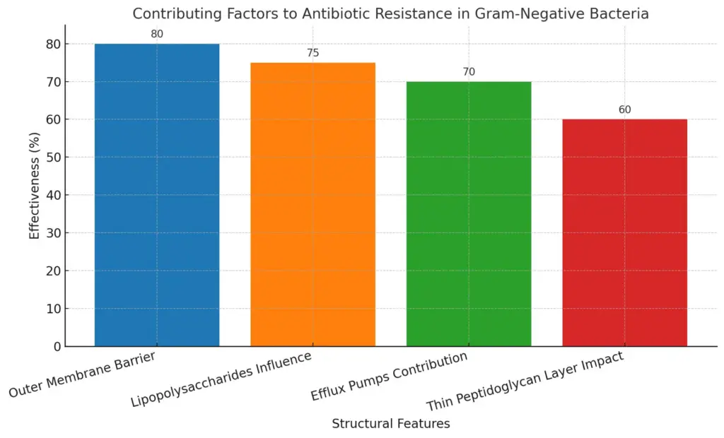
The structural characteristics of Gram-negative bacteria significantly contribute to their heightened antibiotic resistance compared to their Gram-positive counterparts. One of the primary factors is their unique outer membrane, which serves as an effective barrier against many antibiotics, preventing these critical medications from effectively penetrating the bacterial cell. This outer membrane contains lipopolysaccharides, which not only offer protective benefits to the bacteria but also play a significant role in immune evasion, making it harder for the host’s immune system to eliminate the infection. In addition to this formidable barrier, Gram-negative bacteria have evolved to possess efflux pumps that actively expel antimicrobial agents, thereby complicating treatment efforts even further. These efflux pumps are capable of removing a wide variety of drugs from inside the bacterial cell before they can exert their intended effects, leading to increased survival rates for the bacteria. Furthermore, the thin peptidoglycan layer that is located beneath the outer membrane is less accessible to many drugs designed specifically to target bacterial cell walls, which are often more effective against Gram-positive bacteria due to their thicker peptidoglycan layer. Consequently, the combination of these structural defenses creates a multifaceted problem that exacerbates the difficulty in treating infections caused by Gram-negative pathogens. A detailed illustration of this structural difference can enhance understanding of their antibiotic resistance mechanisms, exemplifying why these bacteria represent such a formidable challenge in medical treatment (see image referenced). This complex interplay of structure and function underscores the necessity for ongoing research into effective therapeutic strategies and highlights the critical need for the development of novel antibiotics or alternative treatments tailored specifically to combat these resilient organisms. As science progresses, it becomes increasingly important to stay informed about these challenges in order to innovate new approaches to patient care.

This bar chart illustrates the contributing factors to the antibiotic resistance of Gram-negative bacteria compared to Gram-positive bacteria. Each structural feature is represented by a bar indicating its estimated effectiveness in contributing to antibiotic resistance, with the outer membrane barrier being the most significant at 80%.
| Bacteria | Resistance Rate (%) | Common Antibiotics Affected | Year of Data Collection | Source |
| Escherichia coli | 26 | Ciprofloxacin, Gentamicin, Trimethoprim | 2021 | CDC |
| Klebsiella pneumoniae | 40 | Carbapenems, Cephalosporins | 2021 | CDC |
| Pseudomonas aeruginosa | 32 | Piperacillin/Tazobactam, Meropenem | 2021 | CDC |
| Acinetobacter baumannii | 71 | Colistin, Ampicillin/Sulbactam | 2021 | CDC |
| Salmonella spp. | 23 | Ampicillin, Tetracycline | 2021 | CDC |
Antibiotic Resistance in Gram-Negative Bacteria
IV. The Role of Gram Classification in Medicine
The Gram classification system plays a pivotal role in medical microbiology, primarily by guiding the diagnosis and treatment of bacterial infections. Understanding the fundamental differences between Gram-positive and Gram-negative bacteria helps healthcare professionals make informed decisions regarding antibiotic therapy. For instance, Gram-positive bacteria possess a thick peptidoglycan layer that retains crystal violet dye during the Gram staining process, while Gram-negative bacteria, characterized by a thinner peptidoglycan layer and an outer membrane, do not retain this dye, resulting in a counterstaining with safranin that gives them a pink appearance. This distinction is crucial, as it informs clinicians about the potential antibiotic resistance profiles of the bacteria involved; Gram-negative bacteria often exhibit greater antibiotic resistance due to the presence of efflux pumps and protective outer membranes, which serve as barriers to many commonly used antibiotics. Understanding these characteristics enables healthcare providers to select the most effective treatment options and also highlights the importance of conducting susceptibility testing to guide individualized patient care. Additionally, the structural differences in these bacteria are well-illustrated in studies that emphasize the varying membrane compositions and their implications for treatment strategies. These insights underscore not only the importance of Gram classification in effective medical interventions but also in ongoing research aimed at developing new antibiotics and treatment methodologies that can overcome the challenges posed by antibiotic-resistant bacteria. Ultimately, the Gram classification system remains a vital tool in the ongoing battle against infectious diseases, shaping our approach to clinical care and public health initiatives aimed at controlling bacterial outbreaks.
| Bacteria Type | Common Species | Resistance Rate (%) | Common Antibiotics Resistant |
| Gram-Positive | Staphylococcus aureus | 50 | Methicillin |
| Gram-Positive | Enterococcus faecium | 80 | Vancomycin |
| Gram-Negative | Escherichia coli | 25 | Ciprofloxacin |
| Gram-Negative | Klebsiella pneumoniae | 30 | Carbapenems |
| Gram-Negative | Pseudomonas aeruginosa | 60 | Piperacillin |
Antibiotic Resistance Patterns in Gram-Positive and Gram-Negative Bacteria
A. How Gram Staining Guides Antibiotic Use
Gram staining is a pivotal diagnostic tool that significantly influences antibiotic therapy by distinguishing between Gram-positive and Gram-negative bacteria, each exhibiting unique structural attributes and resistance mechanisms. Gram-positive bacteria possess a thick peptidoglycan layer that retains the crystal violet stain, rendering them susceptible to antibiotics targeting cell wall synthesis, such as penicillin. These characteristics allow clinicians to predict the effectiveness of certain antibiotic treatments, ultimately influencing how patients will be managed in clinical settings. Conversely, Gram-negative bacteria, characterized by their thinner peptidoglycan layer and an external membrane, often evade such treatments and display resistance due to various factors like efflux pumps and porins, as illustrated in [citeX]. The identification of these bacterial types enables healthcare professionals to tailor antibiotic therapy effectively, enhancing treatment outcomes and reducing the risk of resistance. By utilizing Gram staining, physicians can quickly determine whether a bacterial infection is likely caused by Gram-positive or Gram-negative organisms, thereby optimizing their treatment choices. Understanding the structural differences elucidated through Gram staining not only guides clinicians in their selection of appropriate antibiotics but also contributes to broader strategies in managing bacterial infections comprehensively, including efforts in antimicrobial stewardship and reducing unnecessary antibiotic use. Thus, this technique is indispensable in the pursuit of efficient antimicrobial therapy, playing a critical role in the overall framework of infection control and helping to mitigate the growing concern of antibiotic resistance in healthcare settings. The ability to accurately identify bacterial types through Gram staining truly underscores its importance in contemporary medical practice.
| BacteriaType | Antibiotic | Effectiveness | CommonUses |
| Gram-Positive | Penicillin | High | Streptococcus infections, Staphylococcus infections |
| Gram-Positive | Vancomycin | High | Methicillin-resistant Staphylococcus aureus (MRSA) |
| Gram-Negative | Ciprofloxacin | Moderate | Urinary tract infections, respiratory infections |
| Gram-Negative | Aztreonam | High | Infections caused by Pseudomonas aeruginosa |
| Gram-Negative | Gentamicin | High | Severe infections, bloodstream infections |
Antibiotic Efficacy Against Gram-Positive and Gram-Negative Bacteria
B. Challenges in Treating Gram-Negative Infections
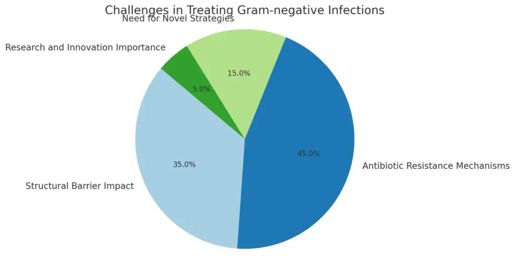
The treatment of Gram-negative infections presents numerous challenges that complicate effective medical intervention, making it a significant hurdle in contemporary medicine. One of the primary difficulties arises from the inherent structural characteristics of Gram-negative bacteria, which are notably distinguished by their unique outer membrane. This outer membrane serves as a formidable barrier to many antibiotics, effectively shielding the bacteria from therapeutic agents that would otherwise compromise their viability. The membrane’s selective permeability is a double-edged sword; it not only limits the entry of helpful antimicrobial agents but also provides formidable protection against external environmental threats, as well as the body’s immune response. Furthermore, the increasing prevalence of antibiotic resistance among Gram-negative pathogens exacerbates the situation significantly. Many strains of these bacteria have successfully developed various sophisticated mechanisms, such as efflux pumps and beta-lactamases, that actively neutralize the effects of commonly used therapeutic agents. This alarming rise in resistance not only undermines existing treatment protocols but also necessitates the urgent exploration of novel antimicrobial strategies, which are crucial for future treatment options. Consequently, the combination of physiological defenses and adaptive resistance mechanisms highlights the critical need for ongoing research and innovation in the development of effective therapies to combat Gram-negative infections. The complexity of these challenges underscores the importance of understanding the fundamental differences between Gram-positive and Gram-negative bacteria in the context of antibiotic resistance. Harnessing this knowledge will be essential for creating targeted solutions and ensuring that healthcare providers are equipped with effective tools to manage resistant infections effectively in the face of an evolving microbial landscape.

This pie chart illustrates the challenges encountered in treating Gram-negative infections. The most significant issue is the antibiotic resistance mechanisms, representing 45% of the challenges, followed by structural barrier impacts at 35%. The necessity for novel strategies and research innovation are acknowledged, at 15% and 5%, respectively.
| Year | Percentage of Infections Resistant to Antibiotics | Common Resistant Strains | Infection Type |
| 2020 | 49.6 | Escherichia coli, Klebsiella pneumoniae | Urinary Tract Infections |
| 2021 | 52.4 | Pseudomonas aeruginosa, Acinetobacter baumannii | Healthcare-associated infections |
| 2022 | 55.3 | Enterobacter cloacae, Salmonella spp. | Bloodstream infections |
| 2023 | 58.1 | Escherichia coli, Klebsiella pneumoniae, Pseudomonas aeruginosa | Respiratory Tract Infections |
Challenges in Treating Gram-Negative Infections
V. Conclusion – Key Differences and Similarities
In summarizing the key differences and similarities between Gram-positive and Gram-negative bacteria, it becomes apparent that their structural variations significantly influence their functions and interactions with antibiotics. Gram-positive bacteria, characterized by a thick peptidoglycan layer and the presence of teichoic acids, typically exhibit a simplified cell structure compared to Gram-negative bacteria, which possess a complex outer membrane and a periplasmic space filled with enzymes and structures that facilitate nutrient transport and defense mechanisms. Notably, this structural distinction impacts their susceptibility to antibiotics; Gram-negative bacteria often demonstrate higher resistance due to their outer membrane acting as a barrier, combined with mechanisms such as efflux pumps that actively expel antimicrobial agents. Despite these differences, both groups share common features, such as the ability to develop resistance through genetic mutations and horizontal gene transfer. Understanding these nuances is critical for developing targeted therapeutic strategies against bacterial infections. The image that most effectively supports this analysis is , which visually delineates the structural contrasts between the two bacterial types, thereby enhancing the discussion on their susceptibility and mechanisms of resistance.

Image : Comparison of Gram-negative and Gram-positive bacterial structures and vesicle types. (The image presents a detailed illustration comparing the structural components of Gram-negative (A) and Gram-positive (B) bacteria. It highlights the Outer Membrane Vesicles (OMVs) in Gram-negative bacteria and Membrane Vesicles (MVs) in Gram-positive bacteria, indicating various biomolecules such as outer membrane proteins, DNA, RNA, peptidoglycan, enzymes, and toxins. The diagram labels key cellular structures including the outer membrane (OM), periplasmic space (PG), and inner membrane (IM) for Gram-negative bacteria, along with lipoteichoic acid (LTA) in Gram-positive bacteria. The visual representation serves to elucidate the differences in bacterial cell wall composition and the types of vesicles produced by these two bacterial groups, which is essential for understanding their biology and pathogenicity.)
| Characteristic | Gram Positive | Gram Negative |
| Cell Wall Structure | Thick peptidoglycan layer | Thin peptidoglycan layer, outer membrane |
| Staining Response | Retains crystal violet stain (appears purple) | Does not retain crystal violet stain (appears pink/red) |
| Sensitivity to Antibiotics | Generally more susceptible to antibiotics such as penicillin | More resistant to antibiotics due to outer membrane |
| Examples | Staphylococcus aureus, Streptococcus pneumoniae | Escherichia coli, Salmonella enterica |
| Toxins Produced | Typically produce exotoxins | Often produce endotoxins |
Key Differences and Similarities of Gram-Positive and Gram-Negative Bacteria
REFERENCES
- Division of Behavioral and Social Sciences and Education. ‘The Prevention and Treatment of Missing Data in Clinical Trials.’ National Research Council, National Academies Press, 12/21/2010
- Kunihiko Nishino. ‘Mechanisms of antibiotic resistance.’ Jun Lin, Frontiers Media SA, 6/1/2015
- Mahendra Rai. ‘Antibiotic Resistance.’ Mechanisms and New Antimicrobial Approaches, Kateryna Kon, Academic Press, 6/14/2016
- Wang Jae Lee. ‘Vitamin C in Human Health and Disease.’ Effects, Mechanisms of Action, and New Guidance on Intake, Springer, 8/6/2019
- Jian-Ying Wang. ‘Regulation of Gastrointestinal Mucosal Growth.’ Second Edition, Rao N. Jaladanki, Biota Publishing, 11/30/2016
- R. Hakenbeck. ‘Bacterial Cell Wall.’ J.-M. Ghuysen, Elsevier, 2/9/1994
- Braj B. Kachru. ‘World Englishes.’ Critical Concepts in Linguistics, Kingsley Bolton, Taylor & Francis, 1/1/2006
- Samuel Reid. ‘Academic Writing Skills 2 Student’s Book.’ Peter Chin, Cambridge University Press, 12/15/2011
- Agency for and Quality. ‘Screening for Methicillin-Resistant Staphylococcus Aureus (Mrsa).’ Comparative Effectiveness Review Number 102, U. S. Department Human Services, CreateSpace Independent Publishing Platform, 8/1/2013
- Alistair McCleery. ‘An Introduction to Book History.’ David Finkelstein, Routledge, 3/13/2006
Image References:
- Image: Schematic representation of bacterial membrane structures and transport mechanisms., Accessed: 2025.https://www.mdpi.com/antibiotics/antibiotics-03-00461/article_deploy/html/images/antibiotics-03-00461-g001.png
- Image: Structural Components of Bacterial Cell Wall, Accessed: 2025.https://pub.mdpi-res.com/molecules/molecules-25-02888/article_deploy/html/images/molecules-25-02888-g001.png?1593341316
- Image: Mechanisms of Antibiotic Resistance in Bacterial Cells, Accessed: 2025.https://media.springernature.com/m685/springer-static/image/art%3A10.1038%2Fs44259-023-00016-1/MediaObjects/44259_2023_16_Fig1_HTML.png
- Image: Comparison of Gram-negative and Gram-positive bacterial structures and vesicle types., Accessed: 2025.https://www.mdpi.com/ijms/ijms-23-11553/article_deploy/html/images/ijms-23-11553-g001.png
