How Do Sponges Digest Food? – (Digestion in Sponges)
Sponges are the simple living multicellular marine-aquatic animals that are found in the coral reefs or in the deep sea water.
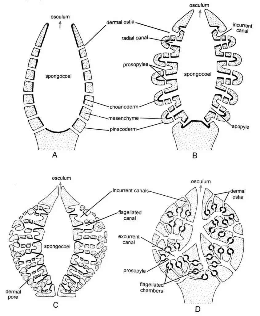
Their body wall is with outer pinacoderm (dermal epithelium), inner choanoderm (gastral epithelium), and gelatinous non-cellular mesenchyme layer in between.
The mesenchyme is actually non-cellular in nature and consists of various other cell types there like Amoebocytes, Scleroblasts, Chromocytes, etc.
The cells lining the canal passage of the sponges help in one way or the other in the proper digestion of the food particles and in the excretion of the wastes.
In order for digestion to take place, the surrounding water has to enter the canal system pathway of the sponge’s body.
The canal system is just the perforation of the body surface by numerous pores for the entry and exit of water into and out of the body of the sponge.
When the water enters the body through the canal pores it brings in food and oxygen into the body and takes out excreta and other reproductive bodies out of the sponge body via. the oscula.

How Do Sponges Digest Food?
STEP 1: Water current enters through porocytes
The body of the sponges is covered externally by pinacoderm which is the outer epidermis layer of cells.
Pinacoderm contains the pinacocytes along with porocytes. The porocytes are actually the pore cells which are special, large, and tubular type in nature.
Porocytes are those tubular cells that make up the pores of a sponge known as Ostia.
Each porocyte contains a central canal-like space communicating with the outside as well as the spongocoel.
Through these pores of the porocytes, water current containing the food particles enter the canal system of the sponges.
STEP 2: Food particles are absorbed by choanocytes
Next the porocytes help in the entry of the water current containing the food particles towards the choanocytes.
Actually, the spongocoel is lined internally by the choanoderm where the choanocytes are located.
Choanocytes are also known as collar cells and they contain a central flagellum, or cilium, surrounded by a collar of microvilli that are connected by a thin membrane.
Choanocytes are used in feeding and for ensuring the flow of water within the animal’s body by the beating of their flagella.
The microvilli of collars act as filter for trapping food particles by engulfing it with the help of pseudopodial action of the choanocytes at the base of the collars.
Resultantly, the food is taken up into the food vacuoles of the choanocytes.
The phase in the food vacuoles is first acidic and then alkaline. And so, here the food particles that were captured undergoes partial digestion.
STEP 3: Amoebocytes receive the food particles from choanocytes
The food particle that was partially digested in the food vacuoles of the choanocytes is now passed on to the wandering amoebocytes in the mesenchyme.
The role of the amoebocytes in the digestion of food in sponges is mainly to deliver the nutrients from choanocytes to other cells within the sponge.
Amoebocytes are also involved in storing the food particles in its food vacuoles for future use. They do also digest some parts of the food for its own use.
A number of enzymes have been isolated from the amoebocytes that include protein, starch, and fat-digestive enzyme.
It is very important to note that both amoebocytes and choanocytes have the ability to transfer food particles to other cells and instead of choanocytes, amoebocytes are the main site of digestion.

Need of Canal System in Digestion
The canal system pathway is the one and only system present in the sponge body that helps it feeding, digestion, transportation of nutrients, excretion, and reproduction as well.
In simple words, its a system of passages connecting various cavities of the sponge body. The water current enters through the pores, passes through the system of canal passages, and circulates inside the body, and exits through the larger pores.
The tiny pores that lead to the canal system are called Ostia. Ostia are those internal pores that internally lead to a system of canals/passages of water and eventually get out to one or larger holes, called oscula. This makes their body canal system work efficiently.
The most vital role in the physiology of sponges is played by the water current flowing in and out of their body through the canal system. The complete life of the sponges depends on their canal system.
Food and oxygen are brought into the body and excreta and reproductive bodies are carried out through the canal system.
The water inside the canal system is caused by the beating of flagella of the collar cells.
The porocytes of the pinacoderm help in the entry of water current carrying the food particles into the canal system. The food containing water current reaches the spongocoel which contains the choanocytes that absorb the food from the water current. Later on, digestion of that captured food is taken care of by the choanocytes and the amoebocytes.
The porocytes form the ostia in the pinacoderm layer, the choanocytes form the gastroderm (choanoderm) inner layer of the spongocoel, and the amoebocytes are present in the mesohyl gelatinous matrix between the pinacoderm and the gastroderm inner layer.
The osculum forms the big pore of the spongocoel through which the water gets out of the sponge’s body.
