Comparative Analysis of Ribosomal Structures: Prokaryotes vs. Eukaryotes
I. Introduction
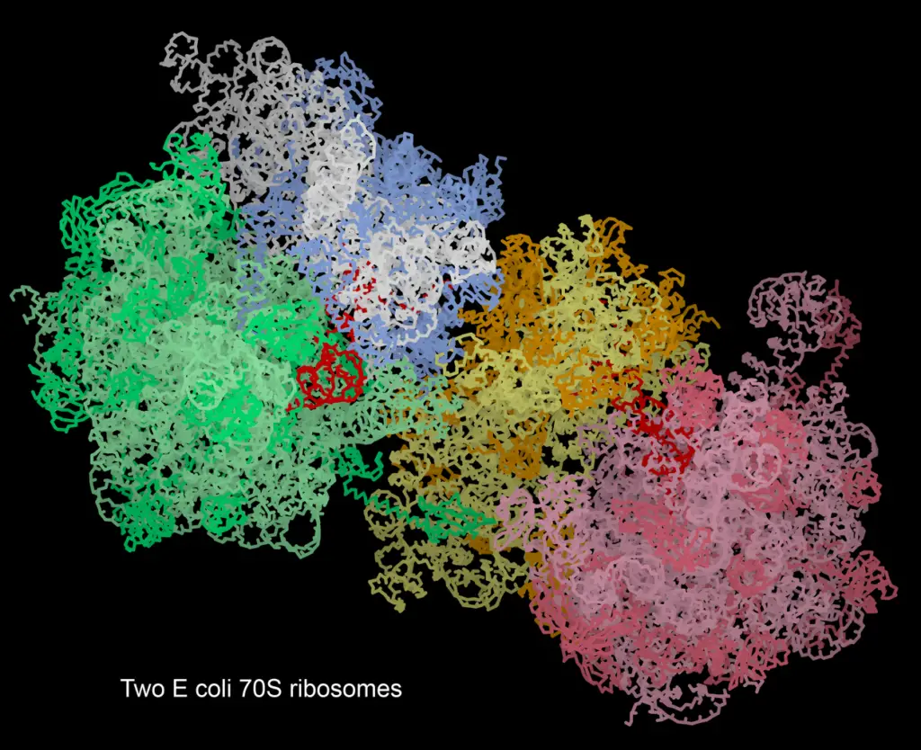
The ribosome is a key cellular machine that makes proteins, showing important differences between prokaryotic and eukaryotic organisms, which highlights their evolutionary and functional differences. Prokaryotic ribosomes are generally smaller and simpler, composed of a 70S subunit that includes a 50S large subunit and a 30S small subunit. Eukaryotic ribosomes, however, are more complex, about 80S in size and made up of a 60S large subunit and a 40S small subunit, reflecting the greater biochemical complexity of eukaryotic systems. Also, the differences in ribosomal RNA (rRNA) and ribosomal proteins (r-proteins) between these two groups are crucial for their protein synthesis processes. Recognizing these differences not only highlights the varied evolutionary journeys of prokaryotes and eukaryotes but also opens the door for a more in-depth study of ribosomal function and its significance for cellular biology. This comparison will carefully look at these structures with a focus on their basic design.

A. Definition and importance of ribosomes in cellular biology
Ribosomes are important machines in cells that help change genetic information into proteins, which is essential for life and cell function. They are made up of ribosomal RNA (rRNA) and proteins, and they have major differences in structure and function between prokaryotes and eukaryotes, affecting evolutionary biology and biotechnology. In prokaryotic cells, ribosomes are found in the cytoplasm and start translating mRNA right after it is made. In contrast, eukaryotic ribosomes are usually attached to the endoplasmic reticulum, which shows that protein synthesis is more complex in these cells. New findings about ribosomes, especially in research on cell-free protein synthesis systems, highlight their important role in biotechnology, improving how proteins are produced and used in various scientific areas (N/A). Additionally, the evolutionary importance of ribosomes is shown in how they serve as basic components of life, reinforcing the link between cellular organisms and viruses as complex biological entities (A Lwoff et al.).
B. Overview of prokaryotic and eukaryotic ribosomal structures
The ribosomal structures in prokaryotes and eukaryotes show key differences that highlight their evolutionary changes and needs. Prokaryotic ribosomes are usually smaller and simpler, made up of a 70S unit that includes a 50S large subunit and a 30S small subunit. This design helps them quickly produce proteins in an efficient way. On the other hand, eukaryotic ribosomes are larger, with an 80S unit formed from a 60S large subunit and a 40S small subunit, which allows for more complex control in the process of translation. The difference in structure also includes the ribosomal RNA (rRNA) and ribosomal proteins (r-proteins) they have; eukaryotic ribosomes contain extra rRNA and a wider range of r-proteins, boosting their functional abilities (Wakely et al.). The evolutionary spread of these ribosomes shows not only their unique biological roles but also the adaptive methods present in both prokaryotic and eukaryotic organisms (Zaeytijd D et al.).
C. Purpose and significance of the comparative analysis
The study of ribosomal structures in prokaryotes and eukaryotes is important for understanding how these key cellular parts have evolved and function. By looking at the similarities and differences in ribosomal design, scientists can learn more about how protein synthesis works in different life forms. This study not only shows how structural changes relate to functional differences but also points out the key ideas of bioinformatics, which have changed the field significantly in the last twenty years by helping to organize and make sense of large amounts of genomic data (Caldwell et al.). Moreover, these studies support a new way of thinking about biological concepts, as discussed in modern biosemiotics, which tries to connect traditional biological findings with more complex views of life processes (Snyder et al.). In the end, this comparative approach deepens our understanding of life’s molecular fabric and sets the stage for progress in biotechnology and medicine.
II. Structural Differences Between Prokaryotic and Eukaryotic Ribosomes
Knowing the differences in structure between prokaryotic and eukaryotic ribosomes is important for understanding their unique roles in protein synthesis. Prokaryotic ribosomes, which have a 70S unit made of 50S and 30S subunits, do not have compartments, letting transcription and translation happen at the same time in the cytoplasm. On the other hand, eukaryotic ribosomes are larger, forming an 80S structure with 60S and 40S subunits, necessitating a more complicated sequence of events, including RNA processing in the nucleus before translation starts in the cytoplasm. Also, the eukaryotic ribosome has more proteins—81 ribosomal proteins compared to fewer in prokaryotes—indicating greater evolutionary complexity and specialization in function (Wakely et al.). Furthermore, prokaryotic ribosomes can quickly adapt to changes in their environment, as seen in the unique features of cereal RIPs in plants, showing their evolution from prokaryotic forms (Zaeytijd D et al.).
A. Size and composition of ribosomal subunits
When looking at the size and make-up of ribosomal subunits, clear differences appear between prokaryotic and eukaryotic structures, showing their evolutionary changes. Prokaryotic ribosomes, like the 70S type, have a 50S large subunit and a 30S small subunit, mainly made from 23S and 5S rRNA in the large subunit and 16S rRNA in the small subunit. On the other hand, eukaryotic ribosomes, such as the 80S type, consist of a 60S large subunit that includes 28S, 5.8S, and 5S rRNA, along with a 40S small subunit that has 18S rRNA. This setup shows a more complicated structure that involves many ribosomal proteins ((Wakely et al.)). This complexity not only boosts their ability to function in gene expression but also gives clues about the evolutionary path that led to the compartmentalization seen in eukaryotic cells ((McCulloch et al.)). Therefore, the size and makeup of ribosomal subunits are key to understanding broader molecular biology and evolution connections.
| Organism | Ribosomal Subunit | Large Subunit | Small Subunit | RRNA Content | Total Proteins |
| Prokaryotes | 70S | 50S | 30S | 5S, 23S, 16S | 55 |
| Eukaryotes | 80S | 60S | 40S | 5S, 28S, 18S | 80 |
Ribosomal Subunits Size and Composition Comparison
B. Differences in rRNA types and their roles
Ribosomal RNA (rRNA) types are key parts of ribosomes and show big differences between prokaryotic and eukaryotic organisms. These differences affect how proteins are made in each type. In prokaryotes, the small ribosomal subunit has 16S rRNA, while the large subunit has 23S and 5S rRNA. This setup allows for a quicker and simpler translation process that happens directly in the cytoplasm. On the other hand, eukaryotic ribosomes are more complex. They have 18S rRNA in the small subunit and 28S, 5.8S, and 5S rRNA in the large subunit, coming together to form the 80S ribosome. This structure is important for various cellular tasks like processing mRNA and building transcription complexes. Additionally, eukaryotes have a nucleus, which adds another level of regulation not seen in prokaryotes. This feature highlights the evolved specialization of rRNA functions in these different organisms (McCulloch et al.), (Wakely et al.).
| Organism Type | RRNA Types | Primary Role | Nucleotide Length (16S) | Nucleotide Length (23S) | Nucleotide Length (5S) | Ribosome Size | |
| Prokaryotes | 16S, 23S, 5S | Protein synthesis and ribosome structure | 1500 | 2900 | 120 | 70S | |
| Eukaryotes | 18S, 28S, 5.8S, 5S | Protein synthesis, ribosome structure and processing | 1800 | 4700 | 160 | 120 | 80S |
Comparative rRNA Types and Their Roles in Prokaryotes and Eukaryotes
C. Variations in ribosomal protein composition
The makeup of ribosomal proteins shows big differences between prokaryotic and eukaryotic systems, showing how they evolved and specialize. Eukaryotic ribosomes have more proteins—81 in plants’ 80S ribosomes—compared to prokaryotes, which usually have about 54 proteins in their 70S ribosomes. Some ribosomal proteins, like RPS15a, have special functions; for example, RPS15aA/F specifically interacts with cytoplasmic rRNA, while RPS15aE is meant for mitochondrial ribosome assembly, showing how this protein family adapted over time (Wakely et al.). Additionally, research shows that many ribosomal proteins have an intrinsic disorder, important for their various roles in assembling and functioning of ribosomes (Dunker et al.). These differences in makeup and roles highlight the complexity and adaptability of ribosomal structures, which are essential for understanding how translation works in different life forms.
| Organism | Ribosomal Proteins | Ribosomal RNA Components | Key Proteins |
| Escherichia coli (Prokaryote) | 54 | 16S rRNA, 23S rRNA, 5S rRNA | S1, S2, S3, S4, S5, S6, S7, S8, S9 |
| Saccharomyces cerevisiae (Yeast, Eukaryote) | 79 | 18S rRNA, 25S rRNA, 5.8S rRNA, 5S rRNA | Rps1A, Rps2, Rps3, Rpl1A, Rpl2A, Rpl3A, Rpl4A, Rpl5 |
| Homo sapiens (Human, Eukaryote) | 82 | 18S rRNA, 28S rRNA, 5.8S rRNA, 5S rRNA | RPS3, RPS5, RPL4, RPL5, RPL7A, RPL11, RPL19 |
Ribosomal Protein Composition in Prokaryotes and Eukaryotes
III. Functional Implications of Ribosomal Structure
The differences in structure of prokaryotic and eukaryotic ribosomes have important effects on how proteins are made. Prokaryotic ribosomes are smaller with a 70S subunit and allow fast translation right in the cytoplasm, which means proteins can be made right after transcription. On the other hand, eukaryotic ribosomes are bigger at 80S and are found in more structured areas, like the rough endoplasmic reticulum. This structure helps control processes related to gene expression and protein development, which is important for the complexity seen in multicellular organisms. Furthermore, the eukaryotic nucleus has added more complexity to how ribosomal structure relates to functions like mRNA processing and stability, as noted in (McCulloch et al.). Research into how ribosomes work, which was recognized with the Nobel Prize in Chemistry 2009, highlights the importance of these structural differences within the larger field of cellular biology and biotechnology.
A. Impact on protein synthesis efficiency
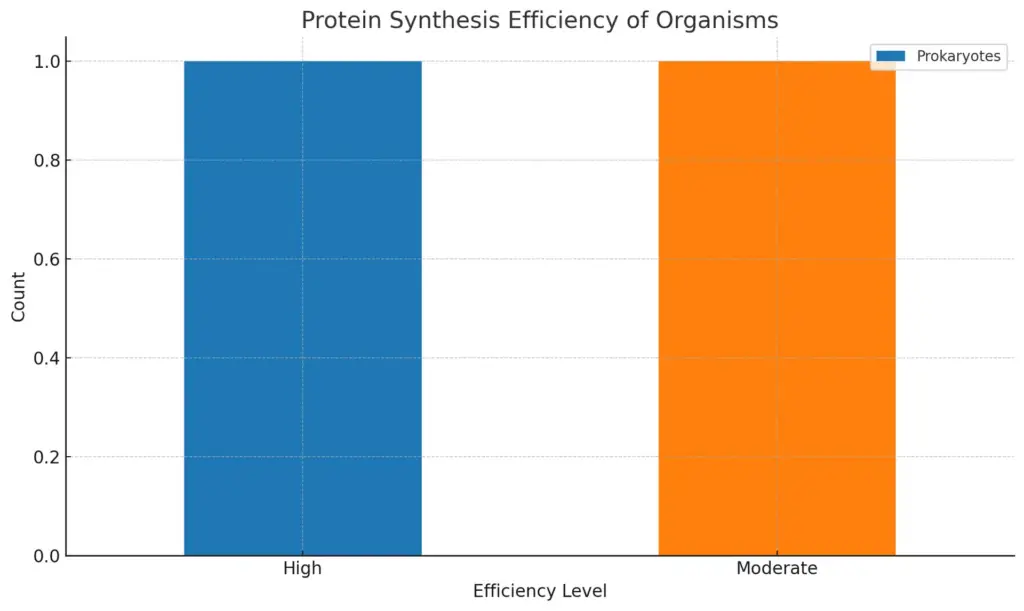
The way protein synthesis works is greatly affected by the differences in structure between prokaryotic and eukaryotic ribosomes, especially in how they signal termination and carry out transcription. In prokaryotes, there are complex ways for starting translation that improve protein diversity and efficiency, showing the importance of riboproteogenomic pipelines to track this variety. The production of different proteoforms from one gene helps prokaryotic organisms adapt to changing environments (Fijalkowska et al.). On the other hand, eukaryotes show more consistent patterns in nucleotide bias near stop codons, which aims to improve translation termination efficiency. Studies show that eukaryotic genomes have steady biases in nucleotide sequences that promote effective termination, leading to high levels of protein production (Cridge et al.). In conclusion, these different molecular strategies illustrate the evolutionary forces that shape ribosomal structure and role in both groups, which directly affects their efficiency in protein synthesis.

The chart displays the comparative protein synthesis efficiency between Prokaryotes and Eukaryotes, illustrating how the two groups differ in their efficiency levels. Prokaryotes show a higher efficiency compared to Eukaryotes, which is classified as moderate.
B. Differences in translation initiation mechanisms
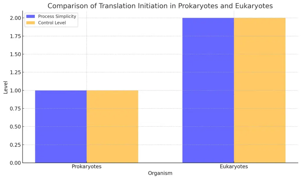
When comparing how prokaryotes and eukaryotes start translation, clear differences show up based on their ribosomes. Prokaryotes use a straightforward method, involving a ribosome binding site called the Shine-Dalgarno sequence. This sequence directly connects with the 16S rRNA in the small subunit of the ribosome, making it easy for the start codon to be recognized efficiently. On the other hand, eukaryotic translation initiation is much more complicated; it requires several initiation factors to bring ribosomal subunits to the cap structure of mRNA, leading to a more controlled process. This added complexity in eukaryotes allows for better regulation of protein synthesis, as seen in the progress of cell-free protein synthesis systems that have come from a better grasp of these processes. Moreover, bioinformatics has greatly changed how we study these mechanisms, providing large databases that help in organizing and understanding extensive genomic information (Caldwell et al.).

The chart compares the translation initiation processes in prokaryotes and eukaryotes by illustrating the simplicity of the processes and the level of control involved. Prokaryotes demonstrate a simple process with low control, while eukaryotes have a more complex process with high control.
C. Role of ribosomal structures in antibiotic susceptibility
The complex structures of ribosomes in prokaryotes and eukaryotes are important for how organisms respond to antibiotics, especially aminoglycosides. Ribosomal RNA (rRNA) forms the main part of where drugs attach, and differences in these attachment sites among ribosomes can greatly change how antibiotics interact with them. Studies show that hybrid ribosomes, which mix eukaryotic rRNA with prokaryotic ribosomes, show increased resistance to some antibiotics. This suggests that changes over time in ribosomal structure have led to different levels of susceptibility (Vasella A et al.). Also, the problem of antibiotic resistance gets worse with practices like using antibiotics in farming, which helps spread resistant bacteria through food, showing how ribosomal structure and antibiotic resistance are linked (FALASCONI et al.). This knowledge is critical for creating effective ways to fight antibiotic resistance in different life forms.
IV. Evolutionary Perspectives on Ribosomal Structures
The differences in ribosomal structures between prokaryotes and eukaryotes offer important understanding of cell complexity and function. Prokaryotic ribosomes are usually smaller and simpler, and they translate genetic information efficiently without the compartmentalization found in eukaryotes. Eukaryotic ribosomes are larger and more organized within the nucleus and cytoplasm. This organization supports a more complex level of gene regulation and processing, as shown by the development of the nuclear envelope and related structures, which improve genome function and expression (McCulloch et al.). Furthermore, the conservation patterns of introns within ribosomal genes among eukaryotic species show a level of complexity that reflects evolutionary changes, highlighting the differences in protein synthesis methods between these groups of life (Hester et al.). Therefore, ribosomal structures not only reveal functional differences between prokaryotic and eukaryotic cells but also represent the broader evolutionary stories that influence biological diversity.
A. Phylogenetic relationships between prokaryotic and eukaryotic ribosomes
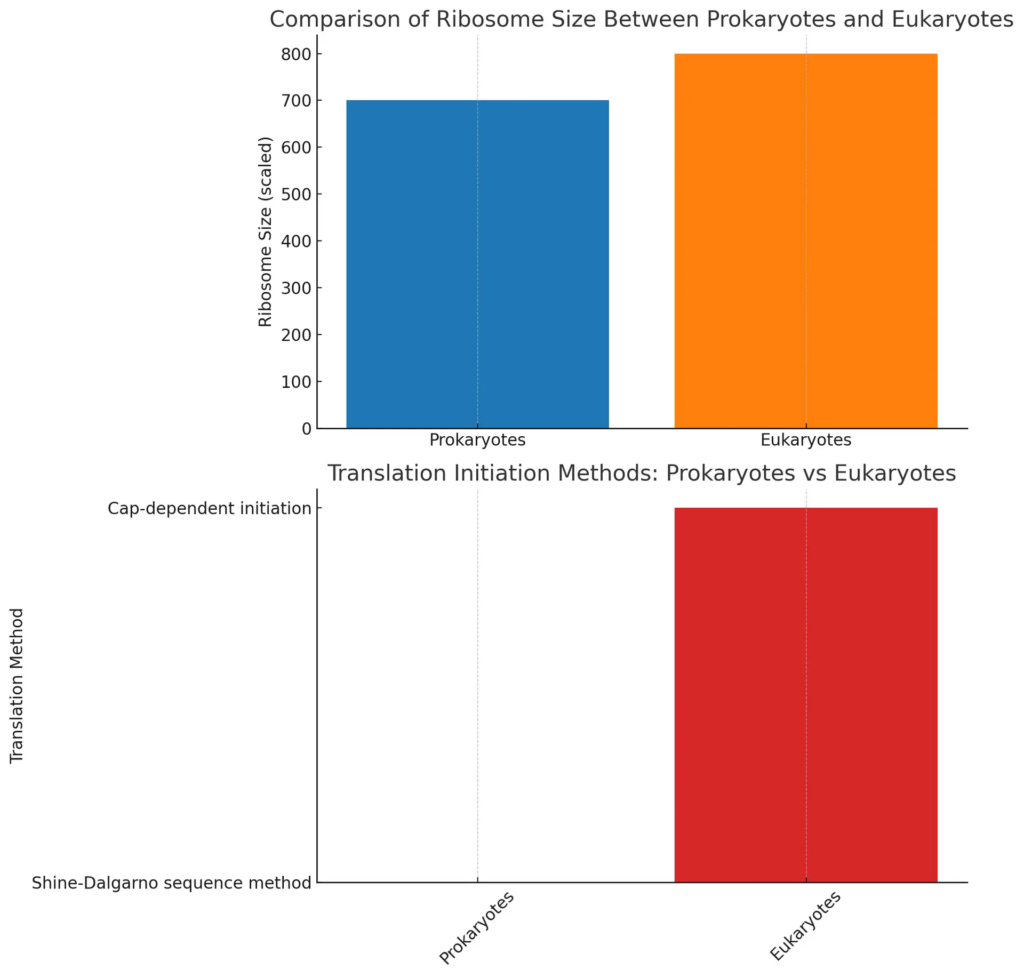
The phylogenetic links between prokaryotic and eukaryotic ribosomes show a complicated history of evolution that highlights how these two life domains diverged. Prokaryotic ribosomes are smaller and simpler than eukaryotic ones, and their core structure mainly comes from archaea, indicating a very old origin that existed before eukaryotes appeared. Recent research suggests that the nucleolus, which is essential for the assembly of eukaryotic ribosomes, probably developed from ancient ribosome assembly processes found in archaea, mixing in factors from bacteria and those specific to eukaryotes to improve its function (Mackowiak et al.). Also, improvements in cell-free protein synthesis (CFPS) techniques have allowed scientists to alter ribosomal functions and production methods on a large scale, showcasing the evolutionary changes that have taken place over time (N/A). Knowing these phylogenetic connections helps us not only understand the structural differences but also reveals important implications for how cells function and evolve among various life forms.

The chart compares ribosome sizes and translation methods used by prokaryotes and eukaryotes. The first graph illustrates the ribosome size, showing that prokaryotes have a 70S ribosome while eukaryotes feature an 80S ribosome, emphasizing the differences in their cellular structures. The second graph presents the methods of translation initiation, highlighting the Shine-Dalgarno sequence method in prokaryotes and cap-dependent initiation in eukaryotes, which are critical processes in protein synthesis.
B. Evolutionary adaptations in ribosomal structures
The changes in ribosomal structures show important differences between prokaryotic and eukaryotic life forms, which reflect their different evolutionary paths. Prokaryotic ribosomes are typically smaller and simpler, which allows for quick protein synthesis in a more variable environment. On the other hand, eukaryotic ribosomes have become larger and more complex, which is necessary for the detailed control of gene expression and cell functions that define eukaryotic cells. These changes not only improve the accuracy of protein translation but also offer an evolutionary benefit by having specialized ribosomal parts that can react to environmental pressures, as indicated in research about natural selection influenced by temperature on genomes and proteomes (Hickey et al.). The background of the differences between prokaryotes and eukaryotes highlights the evolutionary branches that have developed, showing how ribosomal structures have changed to fulfill the unique metabolic needs of these two groups (Sapp J).
C. Implications of ribosomal evolution for understanding cellular complexity
The development of ribosomes is important for grasping how complex cells are, especially when looking at prokaryotic and eukaryotic systems. Eukaryotic ribosomes are more complicated compared to prokaryotic ones and are linked to advanced cellular tasks like compartmentalization and specialized ways of gene expression. The development of the eukaryotic nucleus—seen as a key moment in evolution—allowed for complex regulatory pathways that control transcription and translation, which is quite different from the simpler processes found in prokaryotes. As explained, the nucleus is an important part that not only contains the genetic material but also manages many functions related to DNA maintenance and gene expression controls, highlighting its role in cellular complexity (McCulloch et al.). Along with new technologies like the cell-free protein synthesis system that improve our knowledge of how ribosomes work, these findings highlight the evolutionary importance of ribosomes in forming the complex cellular structures we see now.
V. Conclusion
In conclusion, comparing the ribosomal structures in prokaryotes and eukaryotes shows major differences that indicate their evolutionary changes and functional variations. Prokaryotic ribosomes have a simpler design, allowing for quick protein synthesis directly in the cytoplasm, demonstrating a straightforward process that supports their fast growth and reproduction. On the other hand, eukaryotic ribosomes are more complex, located in the nucleus and affected by various regulatory systems that match the detailed cellular organization seen in these organisms. The development of the nuclear membrane, which did not come from symbiosis, is important in making eukaryotic ribosomal structures different from those in prokaryotes, influencing their roles in keeping the genome stable and in gene expression (McCulloch et al.). Moreover, research on intron conservation in eukaryotic genes highlights the evolutionary pressures that have shaped ribosomal structures, further stressing the distinct nature of eukaryotic ribosomes and their complex biology (Hester et al.).
A. Summary of key findings from the comparative analysis
The comparison of ribosomal structures in prokaryotes and eukaryotes shows important differences that highlight their evolutionary histories and functions. Prokaryotic ribosomes are smaller and have different rRNA and fewer ribosomal proteins, allowing for quick protein synthesis that is important for their simple cellular lives. In contrast, eukaryotic ribosomes are larger and more complex, with extensive rRNA structures and a wider variety of ribosomal proteins, reflecting their more advanced regulatory systems for gene expression and protein translation. These observations are supported by the significant advancements in cell-free protein synthesis (CFPS) technologies, which have proven to be more beneficial in eukaryotic systems, offering reduced sensitivity to product toxicity and higher yields. Additionally, looking at the evolutionary links between archaea and bacteria indicates that the evolution of ribosomes is closely related to wider genomic and functional adaptations in these groups ((Bourne et al.)).
B. Importance of ribosomal structure in biological research
Knowing how ribosomes are built is very important for biology research, especially when looking at differences between prokaryotic and eukaryotic systems. Ribosomes are key players in making proteins, and their different structures show the complex evolutionary changes between these two life forms. Eukaryotic ribosomes are larger and more complex than those in prokaryotes, highlighting advanced roles that support more complicated cellular functions (McCulloch et al.). Additionally, as we examine biological systems through a semiotic lens, the specific features of ribosomes help clarify not just how translation works, but also how genetic expression and regulation happen in various organisms (Snyder et al.). Therefore, studying ribosomal structure is vital for grasping the basic principles of biology and improving our understanding of how cells work and evolve.
C. Future directions for research on ribosomal function and evolution
As research on ribosomal function and evolution continues, future studies will look at the complex links between ribosomal structure and its ability to adapt in various biological situations. One interesting area to explore is the comparison of ribosomes from prokaryotic and eukaryotic organisms, highlighting their evolutionary differences and similarities in response to environmental changes. This could clarify how changes in ribosomes affect the efficiency and accuracy of translation. Additionally, research may concentrate on how ribosome-related factors, like ribosomal proteins and small nucleolar RNAs, help adjust ribosomal function in different physiological settings. Moreover, using advanced imaging methods and cryo-electron tomography might give new understanding of how ribosomes behave during protein synthesis. By investigating these areas, scientists will greatly enhance our knowledge of how ribosomes have evolved and what this means for cellular physiology, thus leading to new biotechnological opportunities.
REFERENCES
- Hickey, Donal A, Singer, Gregory AC. “Genomic and proteomic adaptations to growth at high temperature”. BioMed Central, 2004, https://core.ac.uk/download/pdf/3558987.pdf
- Jan Sapp. “Two faces of the prokaryote concept”. International Microbiology, 2010, https://core.ac.uk/download/159084748.pdf
- De Zaeytijd, Jeroen, Van Damme, Els. “Extensive evolution of cereal ribosome-inactivating proteins translates into unique structural features, activation mechanisms, and physiological roles”. ‘MDPI AG’, 2017, https://core.ac.uk/download/147046433.pdf
- Wakely, Heather. “Binding characteristics and localization of Arabidopsis thaliana ribosomal protein S15a isoforms”. ‘University of Saskatchewan Library’, 2008, https://core.ac.uk/download/226133574.pdf
- McCulloch, Richard, Navarro, Miguel. “The protozoan nucleus”. ‘Elsevier BV’, 2016, https://core.ac.uk/download/77601025.pdf
- Hester, Maria. “Evolution of Gene Structure in Multicellular Eukaryotes”. ScholarWorks@UARK, 2009, https://core.ac.uk/download/84120121.pdf
- Mackowiak, Sebastian, Staub, Eike, Vingron, Martin. “An inventory of yeast proteins associated with nucleolar and ribosomal components”. BioMed Central, 2006, https://core.ac.uk/download/pdf/7539587.pdf
- Dunker, A. Keith, Kurgan, Lukasz, Mizianty, Marcin J., Oldfield, et al.. “A creature with a hundred waggly tails: intrinsically disordered proteins in the ribosome”. ‘Springer Science and Business Media LLC’, 2013, https://core.ac.uk/download/299808966.pdf
- A Lwoff, A Lwoff, B Scola La, B Scola La, C Bandea, CA Suttle, CR Woese, et al.. “Defining Life: The Virus Viewpoint”. Springer Netherlands, 2010, https://core.ac.uk/download/pdf/8388873.pdf
- Caldwell, Rachel Amber. “Investigation of the length distributions of coding and noncoding sequences in relation to gene architecture, function, and expression”. School of Biological Sciences and School of Mathematics and Applied Statistics, 2015, https://core.ac.uk/download/45472448.pdf
- Snyder, Augustus Morrissey. “Biosemiotics as an argument for the recontextualization of biological discoveries: A critical analysis of the biosemiotic model of marcello barbieri.”. JMU Scholarly Commons, 2018, https://core.ac.uk/download/214179343.pdf
- Andrea Vasella, Araujo, Beckers, Beringer, Botero, Campuzano, Carter, et al.. “Engineering the rRNA decoding site of eukaryotic cytosolic ribosomes in bacteria”. Oxford University Press, 2007, https://core.ac.uk/download/pdf/7798465.pdf
- FALASCONI, IRENE. “EVALUATION OF ANTIBIOTIC RESISTANCE PROFILES OF HALOBACTERIA ISOLATED FROM THE FOOD CHAIN”. PIACENZA, 2017, https://core.ac.uk/download/84040026.pdf
- Bourne, Philip E, Valas, Ruben E. “The origin of a derived superkingdom: how a gram-positive bacterium crossed the desert to become an archaeon”. BioMed Central, 2011, https://core.ac.uk/download/pdf/8540486.pdf
- Fijalkowska, Daria, Fijalkowski, Igor, Van Damme, Petra, Willems, et al.. “Bacterial riboproteogenomics : the era of N-terminal proteoform existence revealed”. ‘Oxford University Press (OUP)’, 2020, https://core.ac.uk/download/322827738.pdf
- Cridge, Andrew G., Isaksson, Leif A., Mahagaonkar, Alhad A., Major, et al.. “Comparison of characteristics and function of translation termination signals between and within prokaryotic and eukaryotic organisms”. Oxford University Press, 2006, https://core.ac.uk/download/pdf/3932348.pdf
