Which cell structures are seen in Prokaryotic and Eukaryotic cells? Let’s know in Detail
Cells are the unit of life. No type of life can be believed in existence without the cells.
We all know that cell is the fundamental structure and function of all living organisms on earth, and so anything less than the structure of a cell cannot be considered as independent living, self-sufficient, and self-reliant.
There are two basic types of cells:
- Prokaryotic Cells
- Eukaryotic Cells
What are prokaryotic Cells?
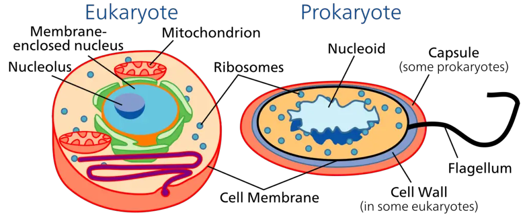
The prokaryotic cells are evolutionary the most primitive type of cells. They do not have a definite true nucleus and so the genetic materials are without an envelope and so the genetic materials remain scattered around in the cytoplasm of the cell.
The size of the cell is between 0.1- 5.0 μm and the organism is unicellular.
The membrane-bound cell organelles are absent and the prokaryotes are usually smaller than eukaryotes.
What are Eukaryotic Cells?
The eukaryotic cells are actually evolved from the prokaryotic cells. They contain organelles like mitochondria bounded by membranes and are located in the cytoplasm.
They have membrane-bound cell organelles and the organisms bearing eukaryotic cells are multicellular in nature.
The nuclear is properly membrane-bound with a nuclear membrane. There is more than one chromosome inside the nucleus.
Cell structures seen in Prokaryotic Cells
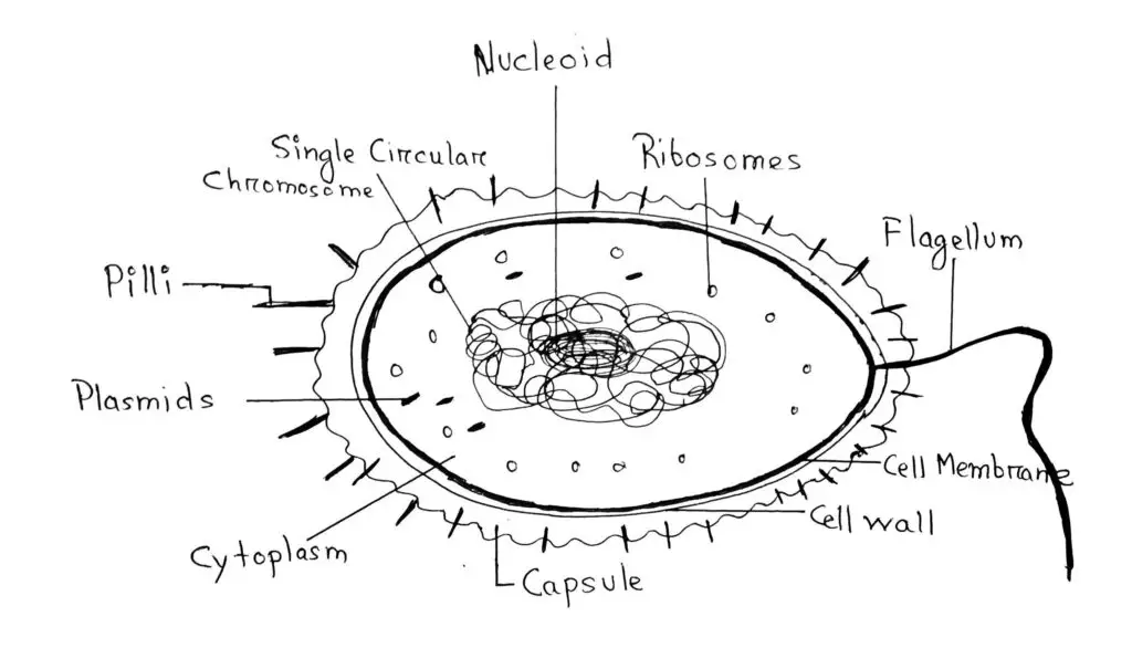
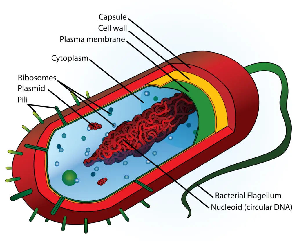
The cell structures that are seen in Prokaryotic Cells are Single Chromosome, Pilus, Falgellum, Cell Envelope, Ribosomes, and Cytoplasm.
1. Single Chromosome
There is a single chromosome that is composed of single double-stranded circular DNA floating freely in the center of the cytoplasm of the cell.
The DNA genetic material is haploid and contains only one copy of the cell. That means that the single chromosome of the cell is haploid.
As the prokaryotes lack a true nucleus so, the chromosome is not contained within a membrane or separated from the rest of the cell. The chromosome is coiled up in a region of the cytoplasm called the nucleoid.
So, the area of the nucleoid region is that area of the cytoplasm that contains the single bacterial DNA molecule.
In the single chromosome of the cytoplasm, the DNA communicates with the cytoplasm and so allowing direct connection to transcription and translation of the cell’s genetic materials.
The Non-essential genes are stored outside of chromosome i.e in the plasmids. The prokaryotic genome is very compact and contains very little non-coding DNA sequences.
2. Pilus
The Pili (Pilus singular) are the small hair-like structures that are seen on the surface of the cell’s outer layer (capsule). It helps attach the prokaryotic bacterial cell to other bacterial cells.
These Pili can have a role in the movement of the cell as well but are more often involved in adherence to surfaces. Shorter pili called fimbriae helps the bacteria attach to surfaces.
The Pili are elongated tubular structures made up of a special Pilin protein. These small structures can be found evenly around the surface of the cell or only on both poles of the cell.
The principle part of the pilus is called the pilus shaft. The pilus shaft is made up of repeating proteins called pilin.
3. Falgellum
Flagella (singular: Flagellum) are used for cell movement by the cell. These are mostly found in the Prokaryotes and only in a very few Eukaryotes.
The prokaryotic flagellum is known to create forward movement using its filament. The filament is corkscrew-shaped that spins to move the organism forward. A prokaryote can have one or several flagella.
Flagella can be found evenly around the surface of the cell or only on both poles of the cell. The bacterial flagellum is made up of a special protein called flagellin.
Its shape is a 20-nanometer-thick hollow tube. It is helical and has a sharp bend just outside the outer membrane; this “hook” allows the axis of the helix to point directly away from the cell with a hook outside to get a hold on.
It is used both as an organ of locomotion and movement as well as a sensory movement. The job of the flagella is like that of a motor boat’s propeller.
Each of the bacteria possesses a certain kind of flagellum makeover with unique arrangements being unique to each bacterium.
As commonly seen in all bacterium that the flagellum rotates and propels the bacterium forward through the surrounding fluid, as the basal body of flagellum acts like a rotary biomolecular motor.
4. Cell Envelope
The cell envelope of the prokaryotes consists of a tightly bound three-layered structure. These 3 layered structure consists of the outer glycocalyx, the middle cell wall, and then the inner plasma membrane.
Although each layer of the envelope performs distinct functions, they act together as a single protective layer.
Glycocalyx: The glycocalyx exists in bacteria as either a capsule or a slime layer. In a capsule, polysaccharides are firmly attached to the cell wall, while in a slime layer, the glycoproteins are loosely attached to the cell wall.
Cell Wall: The cell wall acts as a protective covering providing a strong layer made up of a network of cellulose microfibrils, glycans, pectin, along with lignin biopolymer in some cells. It surrounds the cell body and gives it its shape and rigidity while being located outside of the cell membrane.
Plasma Membrane: The plasma membrane is composed of a semi-fluid lipid bilayer model. It is a thin lipid bilayer (6 to 8 nanometers) that completely surrounds the cell and separates the inside from the outside. It’s non-permeable to ions, proteins, and other molecules, while permeable to other molecules that may move through the membrane.
5. Ribosomes
In prokaryotes, ribosomes are associated with the plasma membrane of the cell. They are about 15 nm by 20 nm in size and are made up of two subunits.
The 2 subunits of Prokaryotic ribosomes are the 50S and 30S subunits, which when present together form the 70S prokaryotic ribosomes.
The 50S subunit contains the 23S and 5S rRNA while the 30S subunit contains the 16S rRNA. Here, S is the Svedberg units.
In total, prokaryotic ribosomes contain around 3 RNA strands along with 52 proteins. Out of these, the 30S ribosome subunit contains around 1 RNA and 21 proteins, while the 50S ribosome subunit contains around 2 RNA and 31 proteins.
6. Cytoplasm
The cytoplasm found in the prokaryotic cells is the same as the cytoplasm in eukaryotic cells. It is a gel-like fluid found bounded by the plasma membrane.
It is a substance in which all of the other cellular components remain suspended.
The prokaryotes have only one membrane i.e the plasma membrane, which encloses the cytoplasm and all of its cellular contents remain suspended in the gel-like fluid.
For unicellular organisms, the cytoplasm is the only place where most of the chemical reactions and metabolic pathways that run the cell take place.
This also means that the cytoplasm houses all the chemicals and components that are used to sustain the life of the cell.
The cytoplasm in both prokaryotic and eukaryotic cells is a viscous liquid with having the same kind of texture and is more kind of gel-like in nature.
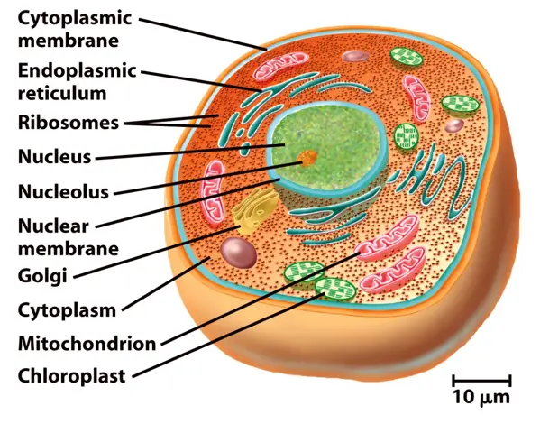
Cell structures seen in Eukaryotic Cells
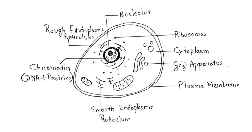
The cell structures that are seen in Eukaryotic Cells are Plasma Membrane, Mitochondria, Endoplasmic Reticulum, Golgi Complex, Nucleus, Cytoplasm, Cytoskeleton, and Microtubules.
1. Plasma Membrane
In animal cells, the Plasma Membrane is the only membrane of the cell. The plant cell has cell wall which the animal cells don’t.
The plasma membrane is semi-permeable in nature and interacts with the outside world. This membrane is similar in structure with that of the prokaryotes.
There in the membrane you will see special extensions in the form of vesicles, tubules, and lamellae extending inwards into the cell from the plasma membrane. These are also called mesosome.
The structure of the plasma membrane shows that it is made up of a phospholipid bilayer with various types of membrane-embedded proteins. The phospholipid bilayer with the various embedded proteins aid in separating the contents that are inside the cell from those that are outside of the cell.
The selectively permeable nature of the plasma membrane allows only a relatively small, non-polar materials to pass through the lipid bilayer either inside or outside of the cell.
Passive, Active, and Osmosis transportation of molecules takes place in and out of the cell through the Plasma membrane only.
2. Mitochondria
Mitochondria (singular: Mitochondrion), are very small in size. These are so small that it can’t be seen under the microscope unless specifically stained.
The main work of the mitochondria is aerobic respiration and the generation of cellular energy.
Mitochondria are small sausage-shaped with diameter of 0.1.0 μm and length of 1.0-4.1 μm.
Each mitochondrion is a double membrane bound organelle in nature. This double membrane bound organelle has its outer membrane and inner membrane dividing its lumen distinctly into two aqueous compartments i.e., the outer compartment and the inner compartment.
The outer membrane is the organelle’s boundary and the inner membrane is the organelle’s matrix.
There in the inner membrane’s inner surface towards the matrix, there are numerous infoldings called the cristae (singular: crista).
3. Endoplasmic Reticulum
The endoplasmic reticulum is the network of tiny tubular structures seen floating in the cytoplasm and often seen with close proximity to the nucleus.
It divides the intracellular space into two parts, luminal which is the space inside the Endoplasmic reticulum, and the extraluminal which is the cytoplasmic space outside the Endoplasmic reticulum.
Endoplasmic Reticulums are of two types: Smooth ER and Rough ER.
Smooth ER: The outer surface of this Endoplasmic Reticulum is smooth with no ribosomes attached to it. These are particularly indulged in the synthesis and secretion of Lipid molecules.
Rough ER: The outer surface of this Endoplasmic Reticulum is rough due to the presence of ribosomes attached to it. These are particularly indulged in the synthesis and secretion of Protein molecules.
4. Golgi Complex / Apparatus
Golgi Apparatus are the site of production of Glycolipids and Glycoproteins.
This cell organelle’s job is to package proteins into vesicles prior to secretion by the cell. It, therefore, plays a key role in the various cell secretory pathways.
These consist of many flat-like, disc-shaped sacs, or cisternae of 0.5 μm to 1.0 μm respectively.
These sacs or cisternae are all situated parallel to each other. A varied number of cisternae are present in a Golgi complex.
5. Nucleus
Nucleus is the brain of the cell. You can call it as the CPU or motherboard of the cell as well.
The Nucleus lies in the center of the cell, having being surrounded by the cytoplasm on all sides.
It’s a double membrane bound structure with small pores in its membrane through which the flow of proteins and ribosomes in and out of the nucleus takes place.
The fluid inside the nucleus is the nucleoplasm. The nuclear matrix inside the nucleoplasm contains nucleolus and chromatin.
There is a structure inside the nucleoplasm which is not membrane-bound and where active ribosomal and RNA synthesis takes place that is the nucleolus.
6. Cytoplasm
Cytoplasm is a thick solution being composed of water, salts, and various proteins that fill each cell and is enclosed by the cell membrane.
The cytoplasm that includes the thick viscous fluid includes all of the material inside the cell and outside of the nucleus.
The organelles that are found in the cytoplasm are mitochondria, Golgi bodies, Endoplasmic Reticulum, Ribosomes, etc.
The composition of cytoplasm includes 85% water, 10-15% proteins, 2-4% lipids, along with nucleic acids, inorganic salts, and polysaccharides in smaller amounts. These things make the cytoplasm thick and highly viscous in texture.
It helps maintain the shape of the cell, its cellular movement, material flow, and exchange in and out of the cell. The cytoplasm makes up 9 out of the 10th of the entire cell.
7. Cytoskeleton
The cytoskeleton is like that of the skeleton of the cell that gives it a firm and a rigid shape.
Cytoskeleton is a network of filaments and tubules that extends throughout a cell from its membrane while passing through the cytoplasm. It is made up of microtubules, actin filaments, and intermediate filaments.
The cytoskeleton supports the cell, gives it shape, and organizes the parts and organelles inside the cell and their locations as well.
They provide a basis for movement and cell division which have significant roles in molecule transport, cell division, and cell signaling as well.
The eukaryotic cytoskeleton consists of three types of filaments microfilaments, intermediate filaments, and microtubules.
These are not other than the elongated chains of proteins.

